About PathFinder
Pathfinder Brain SPECT Imaging, at The Neuroscience Center in Deerfield, IL
High Definition Brain SPECT functional imaging
Director: Dan G. Pavel, M.D.
High Definition Brain SPECT is a non-invasive procedure for functional imaging of the brain (Brain perfusion imaging). With our optimized image acquisition equipment, advanced image processing capabilities and a high quality, detailed and user friendly display of results it has proven a very effective resource for the evaluation of patients with co-existing Neuropsychiatric conditions.
Brain SPECT is performed on the premises of The Neuroscience Center (TNC) in Deerfield, IL, which is a multidisciplinary resource for diagnosis and treatment of complex Neuropsychiatric diseases of youngsters and adults.
The approach at TNC is to integrate the information provided by Brain SPECT in the workup process of refining the differential diagnosis of complex conditions presenting with multiple comorbidities (co-existing conditions) and very often treatment refractory.. Examples of comorbidity can be found in combinations of multiple concussions, concurrent or not, with behavioral changes, developmental illnesses, substance abuse, learning disabilities or cognitive impairment and limbic type seizures. Often patients present after having failed treatment attempts for years. Even in these situations experience has shown that among the multiple treatment plans available at TNC, there is one or a combination of them that ultimately works.
For scheduling : 847.236.9310 / 847.236.9411
Website: www.pathfinder.md
Address :
440 Lake Cook Rd, suite #3
Deerfield, IL 60015
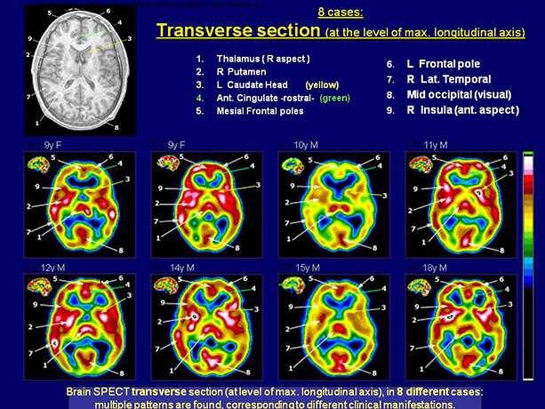
The 8 transverse color slices correspond to 8 different subjects . Nine separate structures are flagged and comparison with their location on an MRI (left upper corner) is also provided . As can be appreciated the same structure can have markedly different levels of perfusion (function) from patient to patient . Take for example arrow # 3 indicating the left caudate head : it shows not only differences between patients but also between right and left . This represents important functional information which cannot be obtained from a standard MRI .